
Tamanho desta previsualização: 509 × 599 pixels. Outras resoluções: 204 × 240 pixels | 626 × 737 pixels.
Imagem numa resolução maior (626 × 737 pixels, tamanho: 27 kB, tipo MIME: image/jpeg)
| Descrição |
English: This scanning electron micrograph (SEM) revealed some of the external ultrastructural details displayed by a flagellated Giardia lamblia protozoan parasite. G. lamblia is the organism responsible for causing the diarrheal disease "giardiasis". Once an animal or person has been infected with this protozoan, the parasite lives in the intestine, and is passed in the stool. Because the parasite is protected by an outer shell, it can survive outside the body, and in the environment for long periods of time.
Cysts are resistant forms and are responsible for transmission of giardiasis. Both cysts and trophozoites can be found in the feces (diagnostic stages). The cysts are hardy and can survive several months in cold water. Infection occurs by the ingestion of cysts in contaminated water, food, or by the fecal-oral route (hands or fomites). In the small intestine, excystation releases trophozoites (each cyst produces two trophozoites). Trophozoites multiply by longitudinal binary fission, remaining in the lumen of the proximal small bowel where they can be free or attached to the mucosa by a ventral sucking disk. Encystation occurs as the parasites transit toward the colon. The cyst is the stage found most commonly in non-diarrheal feces. Because the cysts are infectious when passed in the stool or shortly afterward, person-to-person transmission is possible. While animals are infected with Giardia, their importance as a reservoir is unclear. |
| Fonte | http://phil.cdc.gov/PHIL_Images/8698/8698_lores.jpg |
| Autor | CDC / Mahmud Tari |
| Permissão (Reutilizando este arquivo) |
Copyright Restrictions: None - This image is in the public domain and thus free of any copyright restrictions. As a matter of courtesy we request that the content provider be credited and notified in any public or private usage of this image. |
Esta imagem é um trabalho dos Centers for Disease Control and Prevention, parte do Departamento de Saúde e Serviços Humanos dos Estados Unidos da América, tirada ou feita durante o curso de uma tarefa oficial de um funcionário. Como trabalho do Governo Federal dos Estados Unidos da América, a imagem está no domínio público.
|
|
This media comes from the Centers for Disease Control and Prevention's Public Health Image Library (PHIL), with identification number #8698. Note: Not all PHIL images are public domain; be sure to check copyright status and credit authors and content providers.
|
Histórico do arquivo
Clique em uma data/horário para ver como o arquivo estava em um dado momento.
| Data e horário | Miniatura | Dimensões | Usuário | Comentário | |
|---|---|---|---|---|---|
| atual | 13h31min de 6 de julho de 2022 | | 626 × 737 (27 kB) | wikimediacommons>Chiswick Chap | crop deadspace |
 " class="attachment-atbs-s-4_3 size-atbs-s-4_3 wp-post-image" alt="O que estudar para o enem 2023">
 " class="attachment-atbs-s-4_3 size-atbs-s-4_3 wp-post-image" alt="Qual melhor curso para fazer em 2023">
 " class="attachment-atbs-s-4_3 size-atbs-s-4_3 wp-post-image" alt="Enem: Conteúdos E Aulas On-Line São Opção Para Os Estudantes">
 " class="attachment-atbs-s-4_3 size-atbs-s-4_3 wp-post-image" alt="Como Fazer Uma Carta De Apresentação">
 " class="attachment-atbs-s-4_3 size-atbs-s-4_3 wp-post-image" alt="Como Escrever Uma Boa Redação">
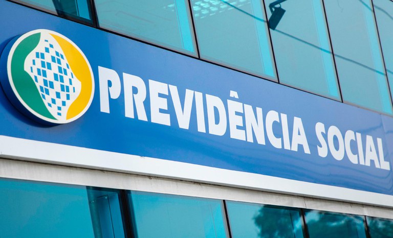 " class="attachment-atbs-s-4_3 size-atbs-s-4_3 wp-post-image" alt="Concurso INSS edital 2022 publicado">

