Amyg.png (189 × 230 pixels, tamanho: 22 kB, tipo MIME: image/png)
Este arquivo é do Wikimedia Commons e pode ser utilizado por outros projetos. Sua página de descrição de arquivo é reproduzida abaixo.
| DescriçãoAmyg.png |
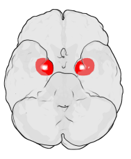
English: Location of the Amygdala in the Human Brain
The figure shows the underside (ventral view) of a semi-transparent human brain, with the front of the brain at the top. The red blobs show the approximate location of the en:amygdala in the en:temporal lobes of the human brain. Note: the amygdala is covered by the ventral temporal cortex (i.e., it is inside the transparent brain). The figure was generated using en:MATLAB and Blender. It is based on MRI imaging data from the Wellcome Department of Imaging Neuroscience, UCL and on amygdalar coordinates from the Talairach brain atlas.Deutsch: Lage der Amygdala im menschlichen Gehirn. Das Bild zeigt eine halbtransparente Abbildung des Gehirns von unten. Das Frontalhirn befindet sich im Bild oben. |
| Fonte |
Originally from en.wikipedia; description page is (was) here * 14:35, 13 February 2004 [[:en:User:Washington irving|Washington irving]] 189×230 (22,159 bytes) <span class="comment">(Location of the Amygdala in the Human Brain)</span> |
| Autor | User Washington irving on en.wikipedia |
| Permissão (Reutilizando este arquivo) |
If you like it, write on my talk page (en:User_talk:Washington_irving). I may be able to add some other brain regions. Released under the GNU Free Documentation License. |

|
Esta imagem (ou todas as imagens neste artigo ou categoria) deveriam ser recriadas usando gráficos vetoriais, como arquivos SVG. Isto tem várias vantagens; veja as imagens para rever para mais informações. Se já criou um arquivo SVG desta imagem, por favor, carregue-o. Depois do novo arquivo SVG ter sido carregado, substitua aqui esta predefinição pela predefinição
{{vector version available|nome da nova imagem.svg}}. |

|
É concedida permissão para copiar, distribuir e/ou modificar este documento nos termos da Licença de Documentação Livre GNU, versão 1.2 ou qualquer versão posterior publicada pela Free Software Foundation; sem Seções Invariantes, sem textos de Capa e sem textos de Contra-Capa. É incluída uma cópia da licença na seção intitulada GNU Free Documentation License.http://www.gnu.org/copyleft/fdl.htmlGFDLGNU Free Documentation Licensetruetrue |
| A utilização deste arquivo é regulada nos termos da licença Creative Commons Atribuição-Partilha nos Termos da Mesma Licença 3.0 Unported. | ||
| ||
| Esta marca de licenciamento foi adicionada a este arquivo durante a atualização da licença GFDL.http://creativecommons.org/licenses/by-sa/3.0/CC BY-SA 3.0Creative Commons Attribution-Share Alike 3.0truetrue |
(Uploaded using CommonsHelper or PushForCommons archive copy at the Wayback Machine)
Legendas
Itens retratados neste arquivo
retrata
image/png
88ba096fb8f571122989c98f3fdca77f84aed3df
22 159 Byte
230 pixel
189 pixel
Histórico do arquivo
Clique em uma data/horário para ver como o arquivo estava em um dado momento.
| Data e horário | Miniatura | Dimensões | Usuário | Comentário | |
|---|---|---|---|---|---|
| atual | 22h31min de 23 de outubro de 2006 |  | 189 × 230 (22 kB) | wikimediacommons>Nicke L | {{Information| |Description= Location of the Amygdala in the Human Brain The figure shows the underside (ventral view) of a semi-transparent human brain, with the front of the brain at the top. The red blobs show the approximate location of the [[:en:amy |
Uso do arquivo
As seguinte página usa este arquivo:
Metadados
Este ficheiro contém informação adicional, provavelmente adicionada a partir da câmara digital ou scanner utilizada para criar ou digitalizar a imagem. Caso o ficheiro tenha sido modificado a partir do seu estado original, alguns detalhes poderão não refletir completamente as mudanças efetuadas.
| Data e hora de modificação do arquivo | 14h43min de 13 de fevereiro de 2004 |
|---|---|
| Resolução horizontal | 28,34 pt/cm |
| Resolução vertical | 28,34 pt/cm |


 " class="attachment-atbs-s-4_3 size-atbs-s-4_3 wp-post-image" alt="O que estudar para o enem 2023">
 " class="attachment-atbs-s-4_3 size-atbs-s-4_3 wp-post-image" alt="Qual melhor curso para fazer em 2023">
 " class="attachment-atbs-s-4_3 size-atbs-s-4_3 wp-post-image" alt="Enem: Conteúdos E Aulas On-Line São Opção Para Os Estudantes">
 " class="attachment-atbs-s-4_3 size-atbs-s-4_3 wp-post-image" alt="Como Fazer Uma Carta De Apresentação">
 " class="attachment-atbs-s-4_3 size-atbs-s-4_3 wp-post-image" alt="Como Escrever Uma Boa Redação">
 " class="attachment-atbs-s-4_3 size-atbs-s-4_3 wp-post-image" alt="Concurso INSS edital 2022 publicado">


